€ 225


Still questions? Contact us
Inner ear anatomy necklace | sterling silver
If you have spent enough time on cochlear physiology, the spiral is the first thing you sketch. The basilar membrane, the organ of Corti, the wave of frequency-specific hair-cell activation flowing from base to apex. Hearing and balance coiled into 35 millimetres of bone, worn here as a 20 mm sterling silver pendant.
The Anatomy and Physiology of the Inner Ear
The inner ear contains two functional systems: the cochlea for hearing and the vestibular apparatus for balance. The cochlea is a spiral bony tube approximately 35 millimetres long, coiled in 2.5 turns within the temporal bone. The basilar membrane runs along its length, increasing in stiffness from base (high frequencies) to apex (low frequencies). Sitting atop the basilar membrane is the organ of Corti, containing approximately 16,000 hair cells arranged in four rows. Sound vibrations cause differential displacement of the basilar membrane along its length, activating different hair cells at different frequencies. Hermann von Helmholtz described the resonance theory in 1863. Georg von Békésy later demonstrated the travelling wave in 1961 (Nobel Prize 1961), showing that basilar membrane displacement propagates along the membrane from base to apex. The vestibular apparatus contains three semicircular canals (anterior, posterior, lateral) plus the utricle and saccule, all filled with endolymph and surrounded by perilymph, detecting head rotation and linear acceleration.
Who Will Recognise It
- audiologists and hearing scientists working on cochlear mechanics and hair-cell physiology
- neuroscientists specializing in vestibular function and balance disorders
- otolaryngologists with a strong interest in inner ear anatomy and physiology
- graduate students whose thesis engages cochlear tonotopy, travelling waves, or vestibular mechanics
The cochlea is the only place where frequency decomposition happens in the body, and anyone who works inside its physics understands why the spiral cannot be simplified.
Explore Related Anatomy Jewelry
- Lungs necklace | silver
- Kidney necklace | silver
- Heart-section necklace | silver
- Uterus necklace | silver
- Knee necklace | silver
FAQ
How does the cochlea convert sound into a neural signal?
Sound waves travel through the external ear canal to the tympanum, which vibrates and transmits vibrations through the three middle ear bones (ossicles) to the oval window at the base of the cochlea. This sets the cochlear fluid (perilymph) in motion. The fluid displacement causes the basilar membrane to vibrate differentially along its length, with different frequencies activating different points. The basilar membrane is stiffest at the base (sensitive to high frequencies) and most compliant at the apex (sensitive to low frequencies). This creates tonotopic organisation, where each hair cell on the organ of Corti responds preferentially to a specific frequency. Hair cells convert basilar membrane displacement into electrical signals that travel along the auditory nerve to the brainstem and cortex.
What is the travelling wave theory?
Georg von Békésy's travelling wave theory (1961, Nobel Prize) explains how the basilar membrane responds to sound. Rather than vibrating uniformly, the basilar membrane exhibits a wave pattern that propagates from the base (stiff) toward the apex (compliant). The peak amplitude of vibration occurs at a specific location along the membrane determined by the sound frequency. High frequencies produce peaks near the base, while low frequencies produce peaks near the apex. This spatial mapping of frequency is the physical basis for tonotopic organisation in the auditory system and explains how the ear separates sound into frequency components.
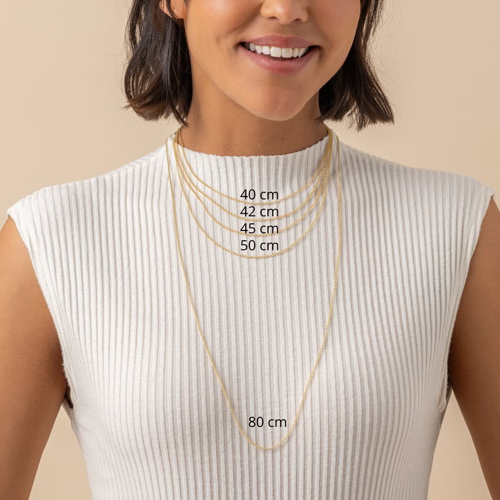
What is the inner ear pendant made of and what chain comes with it?
925 sterling silver, 20 mm pendant on a 45 cm sterling silver chain (ø 1.8 mm) with a 5 cm extender. Nickel-free and hypoallergenic. Free worldwide DHL Express in 1-5 business days, with all import duties and taxes covered. 30-day “Love It or Return It” returns.
Do you have this pendant in gold?
Currently available in sterling silver only. If you are interested in a gold vermeil version, please contact us with a custom order inquiry.

Human Anatomy
Anatomical wonders have never been so elegantly articulated. Our anatomical collection embodies the intricate and awe-inspiring structures that make us who we are. From DNA double helices to neuronal networks, our pieces don't merely imitate—they interpret. The collection serves as a tangible tribute to the hidden beauty within us all, elevating the realms of biology and medicine into wearable art. With exquisite attention to detail, each piece is a dialogue between form and function, revealing the enigmatic eloquence of the human body.


Find your perfect fit: measure an Existing Ring
Finding out your ring size at home is a simple process and can help you shop for jewelry online with confidence.
Necklace length guide